Zero-Radiation Pediatric Ablation Hits 97% Success Using 3D Mapping
Current treatments for cardiac arrhythmias rely mainly on medication or X-ray–guided catheter ablation. For children, however, who are still in critical stages of growth and development, exposure to radiation may pose long-term health risks.
National Taiwan University Children’s Hospital has pioneered the use of three-dimensional (3D) electroanatomical mapping systems to perform fluoroscopy-free catheter ablation. Since treating its first case in 2016, the hospital has completed more than 400 procedures, highest volume in Taiwan, a 97 percent success rate, comparable to conventional X-ray–guided ablation. Average radiation exposure time has been reduced by 98 percent, prompting hospitals in Taiwan and abroad to adopt fluoroscopy-free ablation and opening a new chapter in catheter-based arrhythmia treatment.
The heart’s rhythm depends on its electrical conduction system. A healthy adult heart beats about 60–100 times per minute; children’s heart rates are slightly faster, depending on age. In Taiwan, however, approximately 3 per 1,000 children develop arrhythmias due to abnormalities in this conduction system.
The most common pediatric arrhythmia is paroxysmal supraventricular tachycardia (PSVT), often caused by an extra abnormal conduction pathway between the atria and ventricles, described as an “extra electrical wire.” This can create a short circuit, causing heart rates to suddenly surge to 140–220 beats per minute. More complex arrhythmias may present with rapid or irregular heartbeats, palpitations, dizziness, chest tightness, or chest pain. In severe cases, excessively rapid heart rates can lead to hypotension, fainting, or even life-threatening situations.

Radiation Risks and the Need for Precision
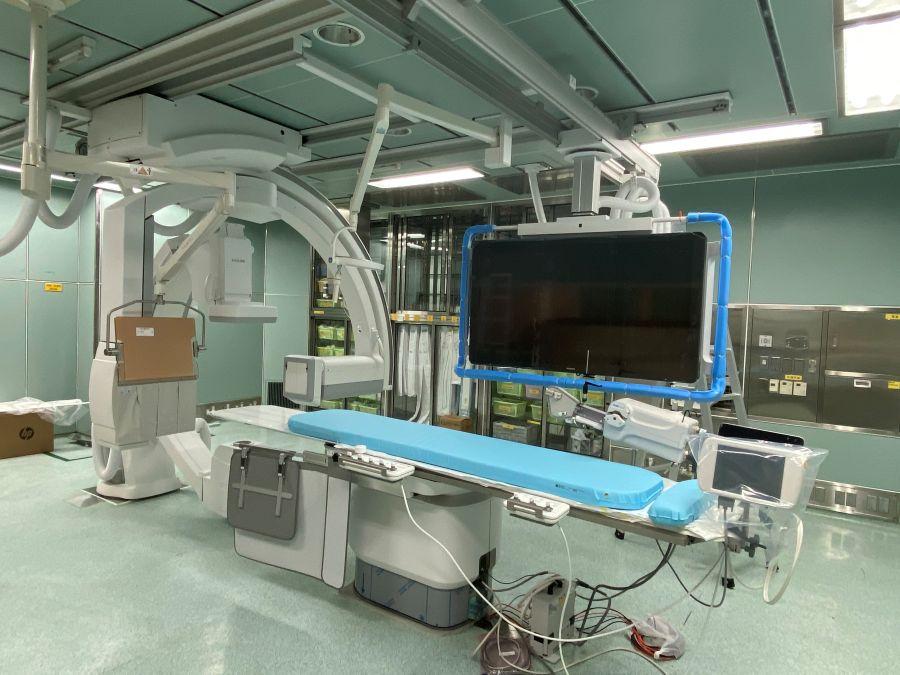
Treating arrhythmias typically involves medication or X-ray–guided catheter ablation. Children’s hearts are smaller and their tissues more delicate, requiring extremely precise localization to avoid damaging healthy structures. Traditionally, X-ray fluoroscopy has been used for guidance, exposing children to radiation during both diagnosis and ablation.
A 2016 report in the European Heart Journal warned that patients undergoing conventional fluoroscopy-guided ablation at age 15 face a 35–40-fold higher lifetime risk of cancer-related death compared with those treated using low-dose fluoroscopy, the urgent need to reduce radiation exposure.
3D Precision Mapping Enables Zero-Radiation Treatment
The idea of minimizing, even eliminating, radiation during pediatric catheter ablation had long been discussed within the NTUH Children’s Hospital team. “In 2016, Professor Ming-Lun Yang, an honorary professor at Miami Children’s Hospital, told us that Europe and the United States had already begun treating arrhythmias with fluoroscopy-free catheter techniques,” said Shun-Nan Chiu, an attending pediatric cardiologist.
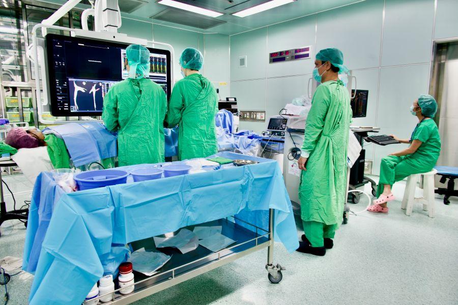
Catheter ablation is an invasive procedure. Cardiologists insert multiple thin catheters through the groin and guide them into the heart. Each catheter is about 100 to 150 centimeters long and roughly the thickness of a pen refill.
A 3D electroanatomical mapping system tracks the catheters in real time using electromagnetic fields. Surface electrodes placed on the child’s skin detect catheter positions. Combined with electrocardiographic analysis and computer processing, the system generates a three-dimensional image of the heart. This allows physicians to precisely target abnormal tissue without X-ray guidance, dramatically reducing radiation exposure.
According to Mei-Huan Wu, former superintendent of NTUH Children’s Hospital and current attending pediatric cardiologist, the system was originally designed to guide complex arrhythmia procedures by providing 3D visualization, the limitations of traditional fluoroscopy, which offers only two-dimensional images. The NTUH team integrated this technology into pediatric catheter labs, leveraging electromagnetic localization to visualize metallic catheters inside the body and, in many cases, achieve completely radiation-free ablation.
Transitioning to a new guidance system was not effortless. Overseas, it typically takes about six months for physicians to adapt from fluoroscopy to 3D mapping. Thanks to extensive clinical experience, the NTUH team mastered the technique in under three months. “Physicians need refined manual skills and prior experience with fluoroscopy-guided ablation,” Chiu noted.
World-First Techniques for Left-Sided Arrhythmias
NTUH Children’s Hospital performed its first fluoroscopy-free pediatric ablation in 2016 and reached a milestone of completely zero-radiation procedures by the fourth case. Right-sided arrhythmias were radiation-free from the outset. Left-sided arrhythmias posed a steeper learning curve because catheters must navigate the aorta and major vessels.
After about 20 cases, the team consistently achieved zero-radiation outcomes. Today, over 97 percent of pediatric patients receive fluoroscopy-free treatment, and simple arrhythmias have been treated entirely without radiation for the past two years.
Internationally, left-sided arrhythmias are often treated via an antegrade approach that punctures the atrial septum, requiring specialized tools and carrying higher procedural risk, often still involving radiation. NTUH Children’s Hospital has pioneered a retrograde, fluoroscopy-free approach via the aorta, now a mature and globally unique technique.
Another major breakthrough is treating infants as young as one year old, weighing as little as 7 kilograms. While medication is typically preferred for children under five, some infants do not respond adequately and require ablation. Because pediatric vessels are extremely small and catheters are generally designed for patients over five years old, the NTUH team minimizes catheter use, employing just two catheters for mapping, marking targets in the 3D system, then switching catheters for ablation—an exceptionally challenging process.
Sharing Innovation, Expanding Impact
Despite initial skepticism, physicians questioned whether zero-radiation procedures were truly safe, the team has actively shared its experience. At the 2018 Asia-Pacific Pediatric Cardiology Congress, colleagues from Japan and Korea openly questioned the feasibility and safety of fluoroscopy-free ablation. Over time, clinical outcomes have answered those doubts.
Building on decades of experience since Taiwan’s first radiofrequency ablation in 1993, NTUH Children’s Hospital has treated the nation’s most complex pediatric arrhythmia cases. Even during the COVID-19 pandemic, the team delivered five international online lectures to share its techniques.
Today, fluoroscopy-free ablation has spread across Taiwan, with hospitals such as Changhua Christian Hospital, Taichung Veterans General Hospital, and National Cheng Kung University Hospital adopting the approach. NTUH Hospital has also applied the technique to pregnant patients.
Recurrence rates after fluoroscopy-free ablation are low, approaching curative outcomes. The next challenge is extending zero-radiation treatment to even more complex cases, including patients with congenital heart disease or implanted pacemakers and defibrillators. While some of these cases still require intracardiac echocardiography and limited radiation, the team plans to address them in future quality-improvement initiatives, working toward safer care for all patients.
Editor’s Note: This article features the Bronze Award recipient of the 25th National Biotechnology and Medical Care Quality Award (Healthcare Institution Category – Featured Medical Services). All titles and positions mentioned reflect the interviewees’ roles at the time of the interviews.